
Julie — l'assistante IA de rédaction radiologique, alignée sur les référentiels ACR/RADS en vigueur — disponible 24h/24.
Téléversez un examen CT, IRM, radiographie, mammographie ou échographie. Julie l'interprète : en moins de 60 secondes, elle rend son interprétation complète sous forme de compte-rendu structuré conforme ACR/RADS avec observations, diagnostics différentiels, score RADS et conduite à tenir — prêt pour votre validation.

Découvrez Julie — votre radiologue IA, en action.
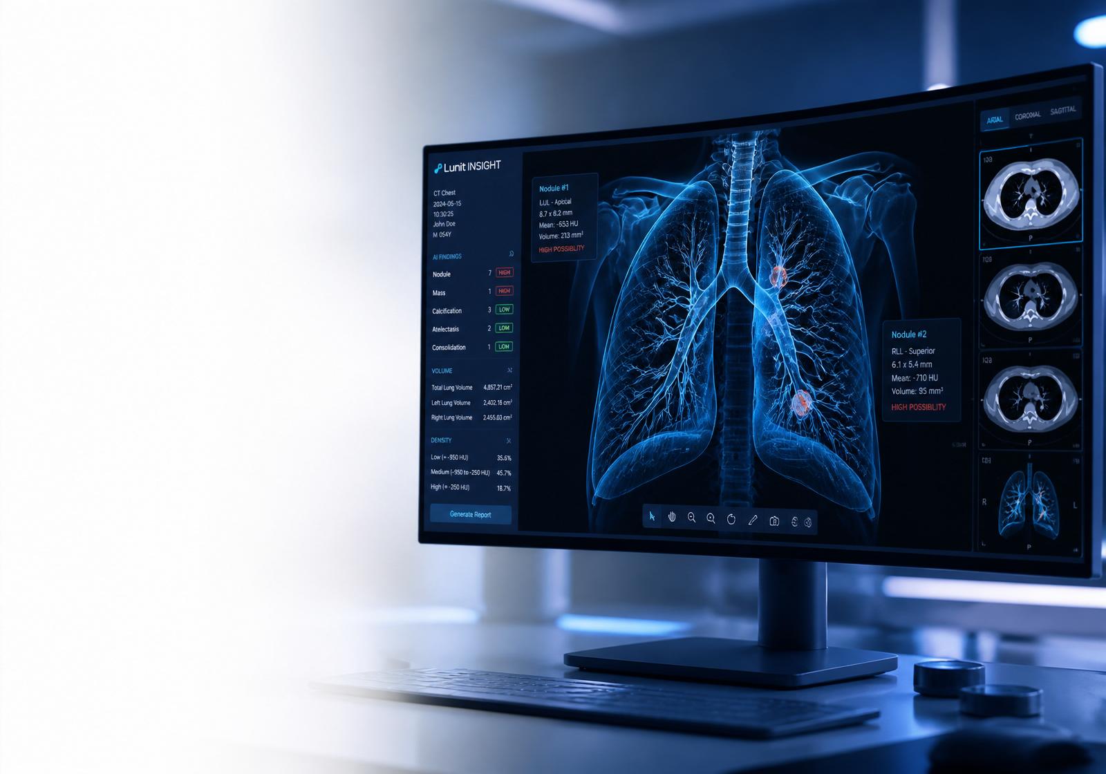
Regardez Julie analyser un scanner thoracique, repérer les anomalies et rédiger un compte rendu structuré — en quelques secondes. Une simulation fidèle de son travail dans la plateforme.
Plus d'examens traités. Moins de temps passé à rédiger. Même contrôle médical.
KANOET augmente votre capacité de lecture sans augmenter votre charge de travail. Pour une clinique : augmentez votre capacité radiologique sans recruter immédiatement. Julie ne pose aucun diagnostic — vous interprétez, vous validez, vous signez.
- Julie01Récupération des antériorités
KANOET rassemble automatiquement les examens précédents du patient et les documents associés.
- Julie02Organisation des informations pertinentes
Contexte clinique, indication, comparatifs et mesures antérieures présentés en un seul écran.
- Radiologue03Interprétation
Le radiologue lit les images et pose l'interprétation. Cette étape reste entièrement humaine.
- Julie04Observations & dictée → compte-rendu
Julie transforme vos observations ou votre dictée en compte-rendu structuré, dans votre style.
- Julie05Impression structurée
Impression hiérarchisée, lexique RADS pertinent et conduite à tenir alignée sur les référentiels.
- Julie06Contrôle de cohérence
Filet de sécurité : omissions, contradictions latéralité/mesures, incohérences observations ↔ impression signalées.
- Radiologue07Validation
Vous acceptez, corrigez ou rejetez chaque proposition. Aucune ligne ne part sans votre accord.
- Radiologue08Compte-rendu final signé
Export PDF / HL7 / FHIR sous votre signature. 100 % de la responsabilité décisionnelle reste au radiologue.
Combien d'examens supplémentaires dans la même journée ?
Les chiffres ci-dessous sont calculés à partir de VOS hypothèses. Nous n'affichons aucune donnée de performance non mesurée chez vous.
À ajuster librement — à valider pendant votre pilote de 14 jours.
Contexte PLFSS 2026 : face à la baisse des tarifs, l'enjeu n'est plus seulement de « gagner du temps », mais de compenser la baisse par un gain de capacité à qualité constante.
Assistant de rédaction non diagnostique — pas de dispositif médical
KANOET n'est pas un dispositif médical au sens du règlement (UE) 2017/745 (MDR) et ne porte pas de marquage CE dispositif médical. Les agents produisent un brouillon de compte rendu et des éléments de relecture ; ils ne posent pas de diagnostic, ne déclenchent aucune décision thérapeutique automatique et ne remplacent aucune lecture médicale.
- Le médecin est l'auteur du compte rendu : rien n'est transmis au dossier patient sans sa validation explicite et tracée.
- Aucune sortie n'est présentée comme un diagnostic, une orientation thérapeutique ou une décision de prise en charge.
- Le périmètre actuel est administratif et rédactionnel ; il ne relève donc pas d'une obligation de marquage CE dispositif médical.
- Toute future fonction à finalité diagnostique ne sera commercialisée qu'après obtention du marquage CE correspondant. Nous n'affichons aucun statut réglementaire tant qu'il n'est pas obtenu.
- La FDA n'est pas pertinente pour le marché France/UE ; elle ne sera invoquée que pour une mise sur le marché américaine effective.
Un workflow de lecture complet, pas un simple classifieur d'images.
Chaque compte-rendu suit une discipline rigoureuse alignée sur les référentiels — templates ACR, scores BI-RADS / LI-RADS, observations structurées, impression et recommandations. Le radiologue garde 100 % de la décision et de la signature.
Lecture multi-modalités
CT, IRM, radiographie, mammographie, échographie, TEP-CT. DICOM et formats standards.
Comptes-rendus structurés ACR/RADS
Scores BI-RADS, LI-RADS, PI-RADS, Lung-RADS appliqués selon la pertinence clinique.
Comparaison avec antériorité
Téléversez un examen antérieur — l'IA signale les lésions nouvelles, résolues ou en progression.
Recherche littérature & recommandations
Diagnostics différentiels ancrés sur PubMed et ACR Appropriateness Criteria.
Dictée vocale
Dictez vos modifications de l'impression et des recommandations à la voix.
Traduction en 12 langues
Traduisez les comptes-rendus complets pour patients internationaux et équipes multi-sites.
Mode Patient
Résumé en langage clair, écrit pour le patient et non pour le radiologue.
Export PDF à votre marque
PDF qualité hôpital avec votre logo et la signature du radiologue.
Lecture urgences & traumatisme
Polytraumatisme relu région par région avec grades AAST, protocoles AVC / embolie / aorte et seuil de communication abaissé pour les trouvailles inattendues.
Mammographie de dépistage (DBT)
Tomosynthèse avec 2D synthétique, catégorie de densité (a–d) rapportée et communiquée, dépistage complémentaire en cas de seins denses.
Téléradiologie & RIS/PACS
Lecture multi-sites avec antériorités et délais de rendu ; les comptes rendus reviennent dans votre RIS/PACS via IHE, HL7 v2 et FHIR R4.
Toujours sous votre validation
Chaque compte-rendu se termine par un avertissement clair — la décision finale vous revient.
Entraînée sur les pathologies que les radiologues lisent réellement chaque jour.
Julie est ajustée sur des millions d'examens publics et dé-identifiés, puis alignée sur les protocoles de lecture ACR/RADS en vigueur. Elle couvre désormais aussi la mammographie de dépistage (BI-RADS), la radiologie d'urgence, la téléradiologie et l'intégration RIS/PACS.
- Thorax : nodules, masses, condensation, pneumothorax, épanchement, embolie
- Neuro : AVC, hémorragie, effet de masse, lésions SEP, schémas d'atrophie
- Abdomen : lésions hépatiques (LI-RADS), masses rénales/surrénales, occlusion
- Musculo-squelettique : fractures, épanchements articulaires, masses des tissus mous
- Sein : caractérisation BI-RADS en mammographie & échographie
- Génito-urinaire : évaluation prostatique PI-RADS en IRM multi-paramétrique
- Mammographie de dépistage : DBT + 2D synthétique, densité (a–d), dépistage complémentaire des seins denses
- Radiologie d'urgence : polytraumatisme par région, AVC / embolie / syndrome aortique, sondes & cathéters, résultats critiques
- Téléradiologie : lecture multi-sites avec antériorités, délais de rendu et remontée des résultats critiques
- Intégration RIS/PACS : commande IHE → worklist DICOM → compte rendu structuré retourné dans votre RIS
Du téléversement au compte-rendu signé en moins de 2 minutes.
- 01
Téléverser l'examen
Glissez un DICOM, JPG ou PNG. Antériorité optionnelle pour comparaison.
- 02
Lecture IA
Protocole aligné référentiels : technique → observations → impression → recommandations.
- 03
Affiner & dicter
Éditez chaque section, dictez à la voix, traduisez, générez le résumé patient.
- 04
Signer & exporter
PDF à votre marque avec votre validation — prêt pour le DPI ou pour le patient.
Chaque étape du protocole de lecture — automatisée, structurée, auditable.
Julie ne « décrit » pas une image. Elle déroule un protocole de lecture complet sur chaque examen, dans l'ordre exact prévu par les référentiels de lecture en vigueur.
1 · Protocole de lecture sur chaque examen
- 01Identifier modalité, région, séquences/phases et adéquation technique
- 02Noter le contexte patient (âge, sexe, question clinique) si fourni
- 03Comparer avec l'antériorité si téléversée — signaler nouveau / résolu / en progression
- 04Revue anatomique systématique (ex. CT thorax : poumons → plèvre → médiastin → vaisseaux → os → étage sus-mésocolique)
- 05Mesurer les lésions en mm, décrire densité / signal / rehaussement, localisation par repères anatomiques
- 06Appliquer le lexique RADS pertinent (BI-RADS, LI-RADS, PI-RADS, Lung-RADS, TI-RADS, O-RADS)
- 07Produire une impression : diagnostics différentiels classés avec niveau de confiance
- 08Recommander la conduite à tenir selon l'ACR Appropriateness Criteria
- 09Terminer chaque compte-rendu par l'avertissement de validation humaine
2 · Sections produites du compte-rendu
3 · Ce qu'elle lit, par modalité
- Pneumonie, condensation, œdème
- Pneumothorax & épanchement
- Nodules / masses
- Cardiomégalie
- Position des sondes & cathéters
- Embolie pulmonaire
- Score nodulaire Lung-RADS
- Pneumopathies interstitielles
- Adénopathies médiastinales
- Pathologie aortique
- AVC ischémique aigu (ASPECTS)
- Classification des hémorragies
- Effet de masse & déviation médiane
- Charge lésionnelle SEP
- Schémas d'atrophie
- Lésions hépatiques LI-RADS
- Masses rénales & surrénales
- Occlusion / ischémie intestinale
- Pancréatite & pathologie canalaire
- Appendicite & diverticulite
- Détection & classification des fractures
- Épanchements articulaires
- Masses des tissus mous
- Tassement & alignement rachidien
- Lésions osseuses (lytiques / condensantes)
- Descripteurs BI-RADS
- Masses vs. calcifications
- Asymétrie & distorsion architecturale
- Catégorie de densité (a–d)
- BI-RADS final 0–6
- Score PI-RADS v2.1
- Zone périphérique vs. transition
- Corrélation DWI / ADC
- Évaluation DCE
- Extension extra-capsulaire
- Score nodulaire TI-RADS
- Composition, échogénicité, marges
- Calcifications
- Évaluation ganglionnaire
- Recommandation cytoponction
- Score O-RADS annexiel
- Évaluation endométriale
- Cartographie des fibromes
- Signes d'endométriose
- Évaluation cervicale
- Polytraumatisme : revue structurée par région avec grades AAST
- Extravasation active & liquide libre signalés comme résultats critiques
- Protocoles AVC (ASPECTS), embolie & dissection aortique
- Sondes, tubes & drains : positionnés ou explicitement rapportés
- Lecture préliminaire vs. finale, avec addenda horodatées
- Tomosynthèse + 2D synthétique
- Voie dépistage vs. diagnostique
- Catégorie de densité (a–d) communiquée à la patiente
- Échographie / IRM complémentaire pour seins denses
- Comparaison aux dépistages antérieurs
4 · Tâches sur chaque compte-rendu
- Comparer avec une antériorité et générer un compte-rendu d'évolution
- Récupérer la preuve PubMed et les critères ACR en ligne
- Dicter à la voix dans n'importe quelle section
- Traduire le compte-rendu complet en 12 langues
- Générer un résumé en mode Patient en langage clair
- Exporter un PDF qualité hôpital avec votre signature
- Piste d'audit de chaque suggestion IA vs. vos modifications
- Acheminer les examens entre sites et retourner le compte rendu signé dans votre RIS/PACS (IHE, HL7, FHIR)
5 · Ce qu'elle ne fera jamais
- Émettre un diagnostic final sans la validation d'un radiologue humain
- Contourner RGPD — chaque examen est chiffré et accès journalisé
- Utiliser vos données patients pour entraîner les modèles de base
- Remplacer un médecin — elle est explicitement un copilote clinique
- Cacher son niveau de confiance — chaque observation est livrée avec sa probabilité
Conçu avec les radiologues, jamais pour les remplacer.
“C'est comme avoir un deuxième regard fiable sur chaque cas complexe — je relis, je décide et je signe.”
“Temps moyen de compte-rendu réduit de 14 à 6 minutes par examen. ROI évident en un mois.”
“Le mode Patient à lui seul a changé notre communication. Les patients comprennent enfin leur imagerie.”